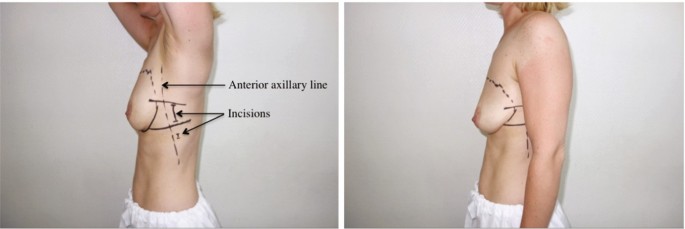
Basic anatomy & Radiology for breast cancer case
By A Mystery Man Writer
Basic anatomy & Radiology for breast cancer case - Download as a PDF or view online for free
This document provides an overview of the anatomy relevant to breast cancer case delineation. It describes the layers of the chest wall including skin, fat, muscles and bones. It outlines the anatomy of structures in the chest including the sternum, ribs, vertebrae, shoulder girdle, and vessels in the neck and chest. The document also details the anatomy of the breast, axilla, supraclavicular fossa, and various muscles of the chest, back, neck and shoulder including the pectoralis major, deltoid, trapezius, and sternocleidomastoid.
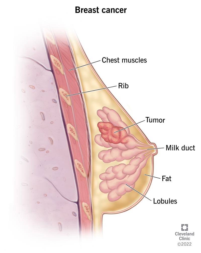
Breast Cancer: Symptoms, Types, Causes & Treatment
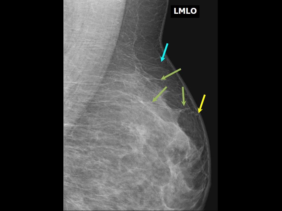
Atlas of breast cancer early detection

Basic anatomy & Radiology for breast cancer case
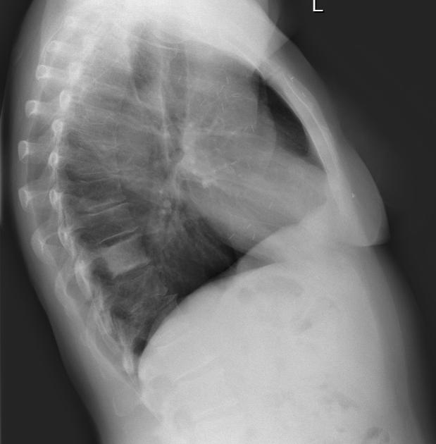
Ivory vertrebra - from breast cancer metastasis, Radiology Case

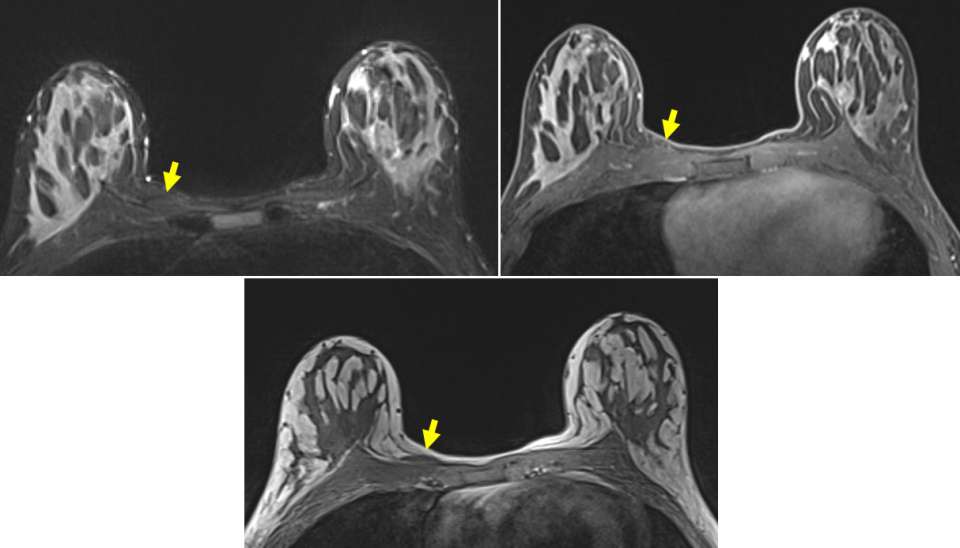
Breast Carcinoma Associated with Poland's Syndrome: One Case Report and Literatures Review
:max_bytes(150000):strip_icc()/1192147_color-5bc5f0c5c9e77c0051fab372.png)
Chest X-Rays and Breast Cancer: Uses, Procedure, Results

Rad Tech CE, ASRT, ARRT® CE, Category A Credits
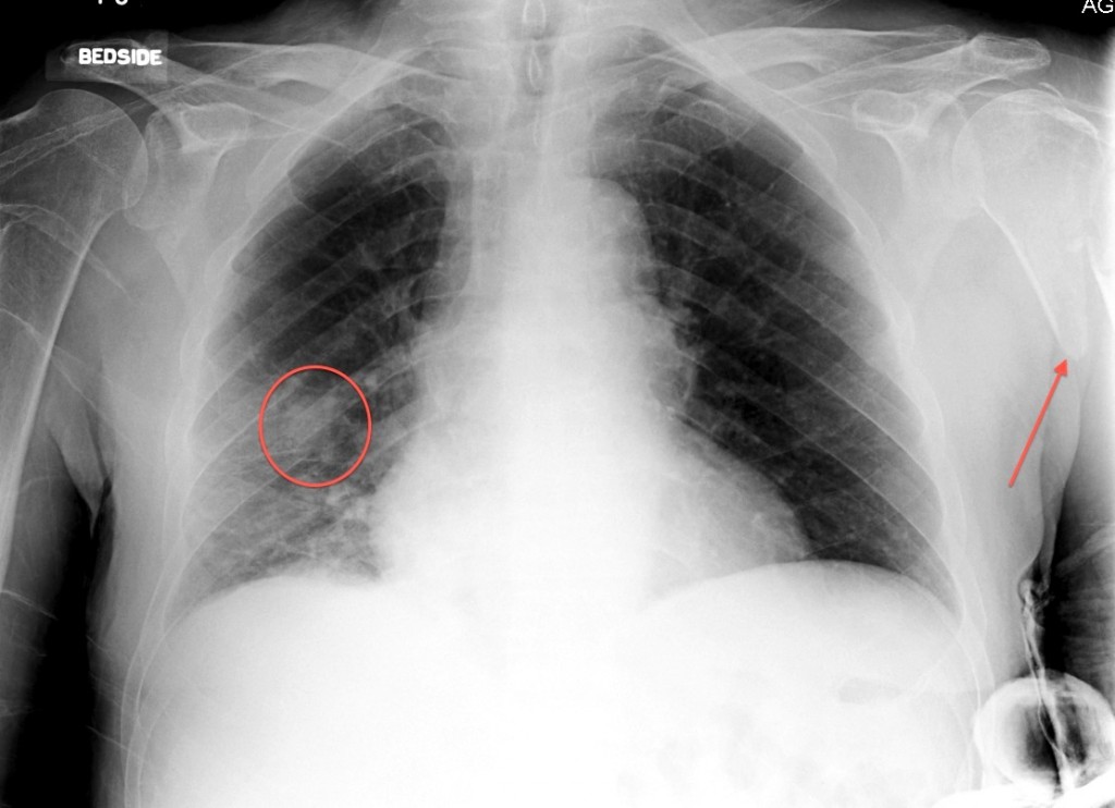
Lung cancer with bone metastasis - Radiology at St. Vincent's University Hospital

Breast ultrasound, Radiology Reference Article
Case: Sternalis Muscle - Radiology
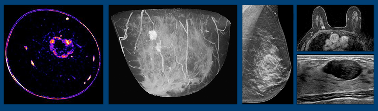
Breast Imaging, Specialties

How A.I. Is Being Used to Detect Cancer That Doctors Miss - The New York Times