Grey scale imaging (ultrasound), Radiology Reference Article

By A Mystery Man Writer
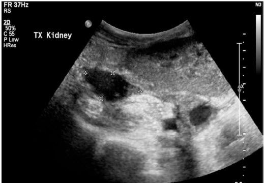
Commonly referred to as B (brightness) mode, the use of grey scale imaging in ultrasound renders a two-dimensional image in which the organs and tissues of interest are depicted as points of v
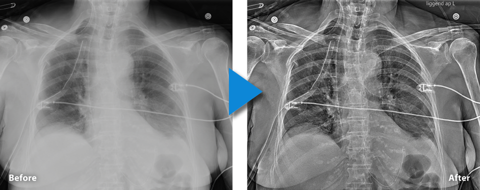
CBr_Mobirex_Plus_Chest1_Catheter_Enhancement_RZ - Canon Medical Switzerland
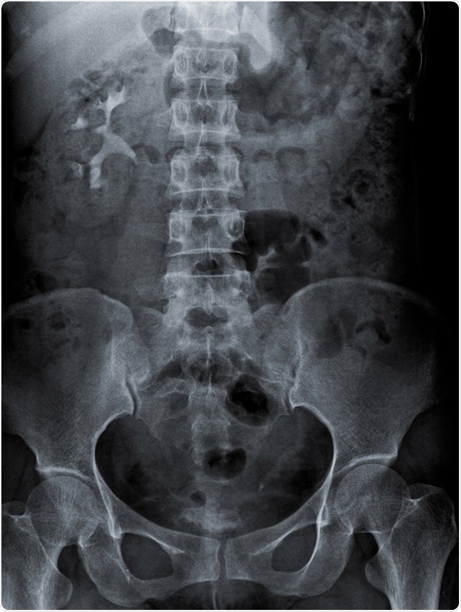
KUB Radiography

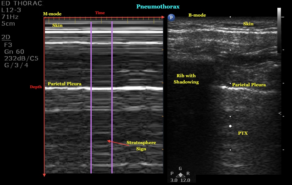
Grayscale Ultrasound Artifacts

PDF] From Grey Scale B-Mode to Elastosonography: Multimodal Ultrasound Imaging in Meningioma Surgery—Pictorial Essay and Literature Review

Multi-contrast submillimetric 3 Tesla hippocampal subfield segmentation protocol and dataset

Animal Models for Human Disorders - Ekam Imaging

Image data of CBTs were collected on gray-scale, CDU, CTA, and MRA. (a)
Primary cerebral cystic echinococcosis in a child from Roman countryside: Source attribution and scoping review of cases from the literature

Posterior Fossa Horns in Hurler Syndrome: Prevalence and Regression