Optical Coherence Tomography: Imaging Mouse Retinal Ganglion Cells In Vivo

By A Mystery Man Writer
Scientific Article | Structural changes in the retina are common manifestations of ophthalmic diseases.
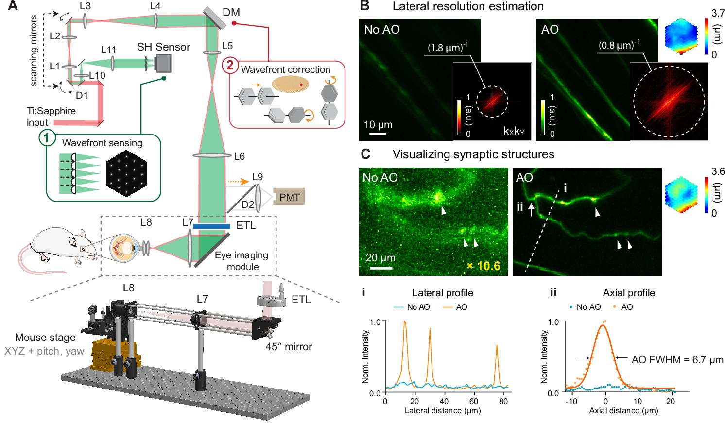
Retinal microvascular and neuronal pathologies probed in vivo by adaptive optical two-photon fluorescence microscopy

Topical Nerve Growth Factor (NGF) restores electrophysiological alterations in the Ins2Akita mouse model of diabetic retinopathy - ScienceDirect

C57Bl/6 retinal scans and posterior segmental layer thi
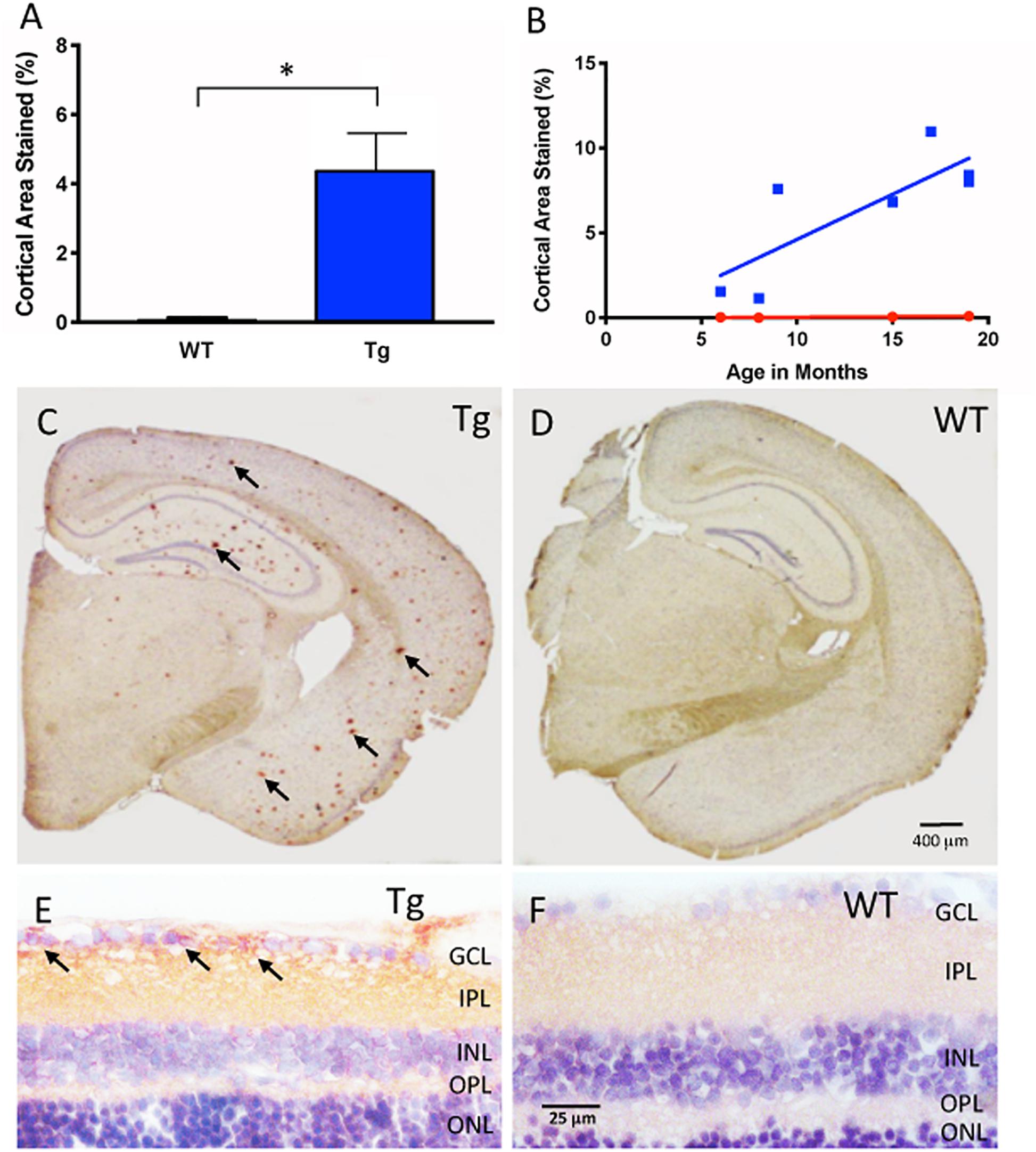
Frontiers In vivo Retinal Fluorescence Imaging With Curcumin in an Alzheimer Mouse Model
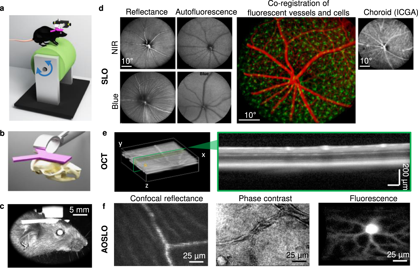
Adaptive-optics SLO imaging combined with widefield OCT and SLO enables precise 3D localization of fluorescent cells in the mouse retina
Applied Sciences, Free Full-Text

All Protocols and Video Articles in JoVE
Spectral-Domain Optical Coherence Tomography of the Rodent Eye
High-resolution structural and functional retinal imaging in the awake behaving mouse

Imaging - Experimental Glaucoma & Imaging Laboratory - Dalhousie University

Transplanted human induced pluripotent stem cells- derived retinal ganglion cells embed within mouse retinas and are electrophysiologically functional - ScienceDirect
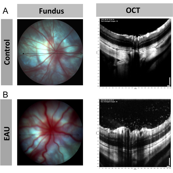
Image-Guided Optical Coherence Tomography to Assess Structural Changes in Rodent Retinas
PDF) Retinal Phenotyping of a Murine Model of Lafora Disease

Imaging - Experimental Glaucoma & Imaging Laboratory - Dalhousie University

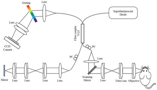
Alignment of Visible-Light Optical Coherence Tomography Fibergrams with Confocal Images of the Same Mouse Retina
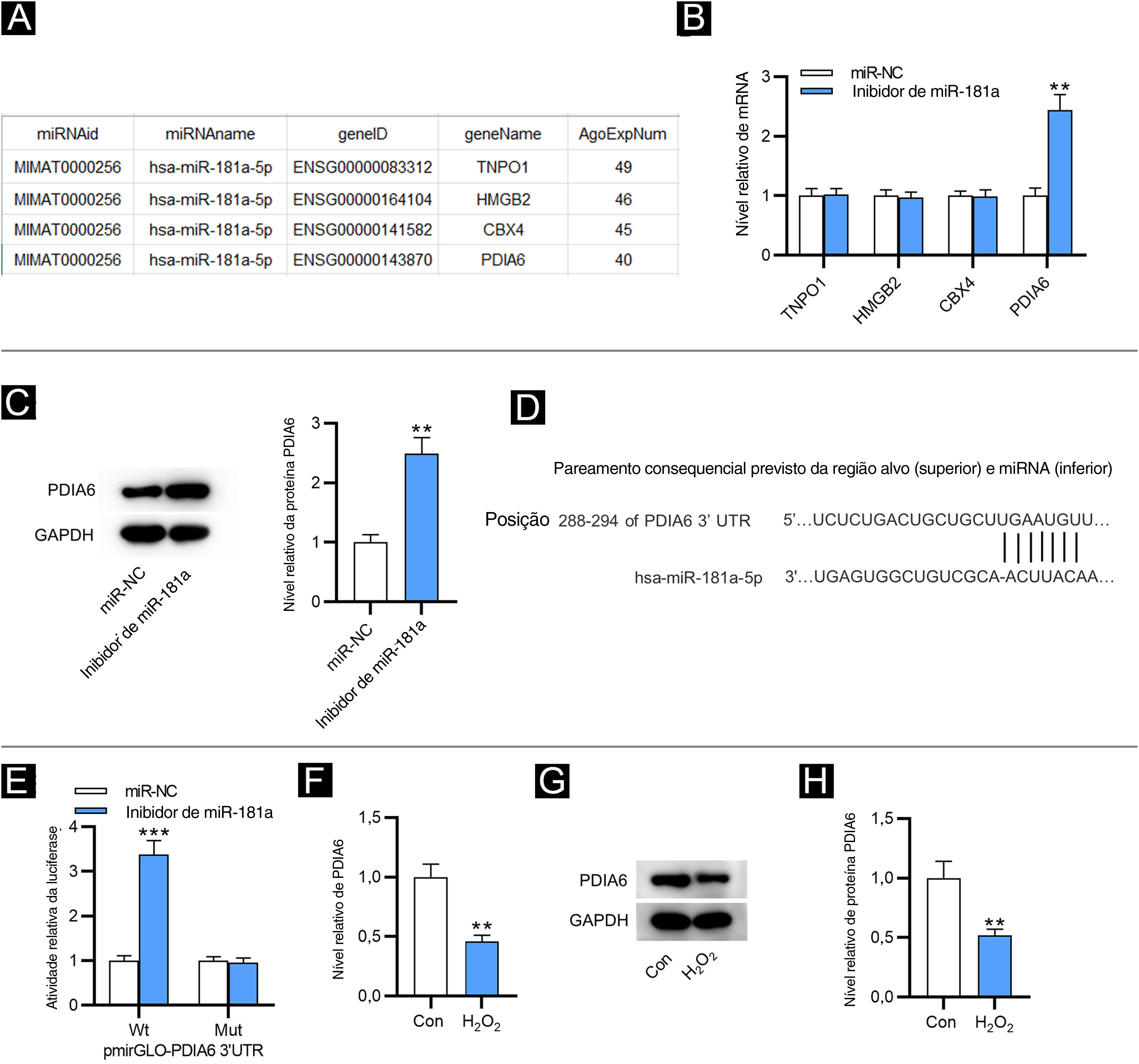
- Sub‐regulação do miR‐181a atenua o estresse oxidativo induzido por H2O2 e a senescência celular atuando sobre PDIA6 em fibroblastos do prepúcio humano
- nanquim ➽ 175 Original artworks, Limited Editions & Prints
- Alinhamento e balanceamento a laser

- Conteúdo - Mentor Odonto

- Mesenchymal stromal cells-based therapy in a murine model of

- Kim Kardashian enlists sports stars for new fashion campaign

- Girlfriend Collective Topanga Halter High Neck Sports Bra Size Medium

- Hanes Originals Women's Mid-Thigh Boxer Brief Underwear
- Nude DD+ Non Pad Plunge Ultimate Comfort Brushed Bra

- Men′ S Sports Sets Summer Short Sleeve Trend Casual Fashion Sport Wear - China Sports Suit and Sport Wear price
